Total Pageviews

Wednesday 11 September 2013

Famous Italian Renaissance Artists and Their Paintings:


Italian Renaissance Artists and Their Paintings:


Animated Clippings of Famous Renaissance Paintings 


Leonardo da Vinci (1452-1519):


Leonardo da Vinci


 Was an Italian Renaissance polymath: painter, sculptor, architect, musician, mathematician, engineer, inventor, anatomist, geologist, cartographer, botanist, and writer. His genius, perhaps more than that of any other figure, epitomized the Renaissance humanist ideal. Leonardo has often been described as the archetype of the Renaissance Man, a man of "unquenchable curiosity" and "feverishly inventive imagination". He is widely considered to be one of the greatest painters of all time and perhaps the most diversely talented person ever to have lived. According to art historian Helen Gardner, the scope and depth of his interests were without precedent and "his mind and personality seem to us superhuman, the man himself mysterious and remote". Marco Rosci states that while there is much speculation about Leonardo, his vision of the world is essentially logical rather than mysterious, and that the empirical methods he employed were unusual for his time.

Mona_Lisa


DaVinci_Last-Supper


Michelangelo Buonarroti (1475-1564)


Portrait of Michelangelo


commonly known as Michelangelo was an Italian sculptor, painter, architect, poet, and engineer of the High Renaissance who exerted an unparalleled influence on the development of Western art. Despite making few forays beyond the arts, his versatility in the disciplines he took up was of such a high order that he is often considered a contender for the title of the archetypal Renaissance man, along with his fellow Italian Leonardo da Vinci.

The-Torment-of-Saint-Anthony-by-Michelangelo.


The-Conversion-of-Saul-by-Michelangelo


Raphael Sanzio (1483-1520):


Raphael


better known simply as Raphael, was an Italian painter and architect of the High Renaissance. His work is admired for its clarity of form and ease of composition and for its visual achievement of the Neoplatonic ideal of human grandeur. Together with Michelangelo and Leonardo da Vinci, he forms the traditional trinity of great masters of that period.

Raphael_-_Resurrection_of_Christ


School-of-athens-by-raphael.


Sandro Botticelli (1445-1510):


Probable self-portrait of Botticelli, in his Adoration of the Magi.


was an Italian painter of the Early Renaissance. He belonged to the Florentine school under the patronage of Lorenzo de' Medici, a movement that Giorgio Vasari would characterize less than a hundred years later as a "golden age", a thought, suitably enough, he expressed at the head of his Vita of Botticelli. Botticelli's posthumous reputation suffered until the late 19th century; since then his work has been seen to represent the linear grace of Early Renaissance painting. Among his best known works are The Birth of Venus and Primavera.


Adoration-of-the-Magi-by-Sandro-Botticelli.

File:Sandro Botticelli - La nascita di Venere - Google Art Project - edited.jpg
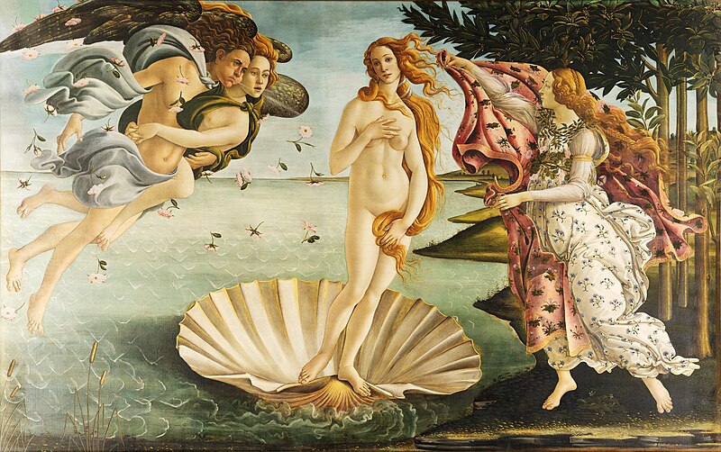
Birth-of-Venus-by-Sandro_Botticelli


Titian (1488-1576):


Self-Portrait of Titian


was an Italian painter, the most important member of the 16th-century Venetian school. He was born in Pieve di Cadore, near Belluno (in Veneto), in the Republic of Venice. During his lifetime he was often called da Cadore, taken from the place of his birth.Recognized by his contemporaries as "The Sun Amidst Small Stars" (recalling the famous final line of Dante's Paradiso), Titian was one of the most versatile of Italian painters, equally adept with portraits, landscape backgrounds, and mythological and religious subjects. His painting methods, particularly in the application and use of color, would exercise a profound influence not only on painters of the Italian Renaissance, but on future generations of Western art

Emperor-Charles-by-Titian


The-Worship-of-Venus-by-Titian

Albrecht Durer (1471-1528):


Durer_selfporitrait


was a German painter, engraver, printmaker, mathematician, and theorist from Nuremberg. His high-quality woodcuts (nowadays often called Meisterstiche or "master prints") established his reputation and influence across Europe when he was still in his twenties, and he has been conventionally regarded as the greatest artist of the Northern Renaissance ever since. His vast body of work includes altarpieces, religious works, numerous portraits and self-portraits, and copper engravings. The woodcuts, such as the Apocalypse series (1498), retain a more Gothic flavour than the rest of his work. His well-known prints include the Knight, Death, and the Devil (1513), Saint Jerome in his Study (1514) and Melencolia I (1514), which has been the subject of extensive analysis and interpretation. His watercolours also mark him as one of the first European landscape artists, while his ambitious woodcuts revolutionized the potential of that medium.


Albrecht_Durer-St_Jerome_in_the_Wilderness-1495


Albrecht_Dürer_adam_and_eve.


Wednesday 27 February 2013

My Unusual Experiment with Mobile Phone`s Camera on My Micro Microscope


My Microscopic Slides Display In Animated Form


My Micro Microscope


My Unusual Experiment with Mobile Phone`s Camera on  My Micro Microscope


This Low Cost Beautiful Microscope Manufactured by  Micro Instruments. This Microscope is very low weight  handy can be carry anywhere. Battery Operated Internal Light installed therefore no external light Needed.This Microscope is suitable for any College Level Biology Students. Magnification as good as any other Normal Compound  Microscope.


My Micro Microscope


Specification of This Microscope is Given Below:

Model MM 02

Magnification : 100x, 450x (Using 10x Eye Pc) : 150x, 675x (Using 15x Eye Pc.)

Use its own Light Source (Battery Operated)

Battery : 9V, 6F 22 - One Required

Completely New and Very Special objectives have been used, Patent Technology

Viewing Field Same as Compound Microscope.

Weight Less than 200gms.

Can Be used Day and Night
                             
I Have Taken those Pictures (Given Below) With My LG Mobile Phone having 2 Megapixel Camera On  My Micro Microscope. 


Bacteria-Coccus-Form Overall Distant View


1) Bacteria-Coccus-Form:


Bacteria-Coccus-Form Close View


2) Lung-Mammal:


Mammalian Lung Picture Taken By Me


3) Bone-Mammal:


Bone Picture Taken By Me


4) Small-Intestine-Mammal:


Mammalian Small Intestine Picture Taken By Me


Mammalian Small Intestine Picture From Internet


5) Kidney-Mammal:

Mammalian Kidney Picture taken By Me


Mammalian Kidney Picture from Internet


6) Heart- Mammal:


Mammalian Heart picture Taken By me


7) Pancreas:


Pancreas Picture Taken By Me


Pancreas Picture with its Parts From Internet


1 - acinus
2 - islet of Langerhans
3 - interlobular connective tissue septa
4 - intralobular duct
5 - interlobular duct


8) Blood-Human:


Human Blood Picture Taken By Me


9) Plant Cells:


Plant Cell Picture Taken By Me


10) Monocot- Leaf:


Monocot Leaf Picture Taken By Me


Monocot Leaf Picture with its Part from Internet


11) Monocot-Stem:


Monocot Stem Picture Taken By Me


Monocot Stem Picture with its Parts from Internet


12) Dicot- Leaf:


Dicot Leaf  close Picture taken by Me


Dicot Leaf  Overall Distant Picture taken by Me


Dicot Leaf  Overall Distant Picture with its parts from Internet


Human Male Reproductive Organs

Male Reproductive Organs in Animated Form


All Male Reproductive Organs in a Single Picture


REPRODUCTIVE SYSTEM:
The ability to reproduce is one if the properties distinguishing living from non-living matter.The mo0re primitive the animal,simpler the process of reproduction.In human being the process is one of  sexual reproduction,in which the male and female organs differ anatomically and physiologically.
  
 Both males and females produce specialized reproductive germ cell, called gametes. The male gametes are called spermatozoa and female gametes are called ova. They contain the genetic material, or genes called chromosomes,which pass inherited characteristics on to the next generation. Other body cell possess 46 chromosomes arranged in 23 pairs but the gametes contain only 23, one from each pair.Gametes are formed by meiosis . At fertilisation,the fusion of an ovum and a spermatozoon,the resulting cell is called zygote, and now possesses the full complement of 46 chromosomes.The zygote embeds itself in the wall of the uterus where it grows and develop during the 40-weeks gestation period before birth.


Male Reproductive Organs


The functions of male reproductive system are: 


1) Production of spermatozoa.
2) Transmission of spermatozoa to the female.

URETHRA:
The male urethra provides a common pathway for the flow of urine and semen, the combined secretions of the male reproductive organs.It is about 19 to 20 cm long and consists of three parts.The prostatic urethra originates at the urethral orifies of the bladder and passes through the prostate gland.The membranous urethra is the shortest and narrowest part and extends from the prostate gland to the bulb of the penis, after passing through the perineal membrane.The spongiose or penile urethra lies within the corpus spongiosum of the penis and terminates at the external urethral orifies in the glans penis.


Penis, Urethra and other Male reproductive Organs


PENIS:

The penis has a root and a body .The root lies in the perineum and the body surrounds the urethra. It is formed by three cylindrical masses of erectile tissue and smooth muscle.The erectile tissue is supported by fibrous tissue and covered with skin and has a rich blood supply.The 2 lateral columns are called the corpora cavernosa and the column between them, containing the urethra,is the corpus spongiosum . At its tip it is expanded into a triangular structure known as the glans penis.Just above the glans the skin is folded upon itself and forms a movable double layer, The foreskin or prepuce.

Arterial blood is supplied by deep dorsal and bulbar arteries of the penis, which are branches from the internal pudendal arteries. A series of veins drain blood to the internal pudendal and internal iliac veins.The penis is supplied by autonomic and somatic nerves.Parasympathetic stimulation leads to  filling of spongy erectile tissue with blood,caused by arteriolar dilatation and venoconstiction, which increases blood flow into the penis and obstructs outflow. The penis therefore becomes engorged and erect, essential for intercourse.


Internal Structure of Testis


TESTES:
The testes are the reproductive glands of the male and are the equivalent of the ovaries in the female.They are about 4.5cm long, 2.5 cm wide and 3 cm thick and are suspended in the scrotum by the spermatic cords.They are surrounded by 3 layers of tissue.

Tunica vaginalis:
This is a double membrane, forming the outer covering of the testes,and is down growth of the abdominal and pelvic peritoneum.During early fetal life, the testes, and is down growth of the abdominal and pelvic peritoneum.

Tunica albuginea:
This is a fibrous covering beneath the tunica vaginalis that surrounds the testes.In growths form septa, dividing the glandular structure of the testes into lobules.

Tunica vasculosa:
This consists of a network of capillaries supported by delicate connective tissue.

Function:
Spermatozoa(sperm) are produced in the seminiferous tubules of the testes, and mature as they pass through the long and convoluted epididymis, where they are stored.The hormone controls the sperm production is FSH from anterior pituitory.A mature sperm has a head , a body and long whip like tail used for motility. The head is almost completely filled by nucleus, containing its DNA.It also contain the enzymes that required to penetrate The outer layers of ovum to reach and fused with its nucleus. The body of the sperm is packed with mitochondria, to fuel the propelling action of the tail that powers the sperm along the female reproductive tract.Successful spermatogenesis takes place at a temperature about 3 degree below the normal temperature.The testes are cooledby their position outside the abdominal cavity,and the thin outer covering of the scrotum has very little insulating fat.


Penis, Urethra and other Male reproductive Organs


PROSTATE GLAND:
This lies in the pelvic cavity in front of the rectum and behind the symphysis pubis, surrounding the first part of the urethra.It consists of an outer covering, a layer of smooth muscle and glandular substance composed of columnar epithelium cells.It secrete a thin, milky fluid that makes up about 30 % of semen , and give its milky appearance.It contains a clotting enzymes, which thickens the semen in the vagina, increasing the likelihood of the semen being retained close to the cervix.

SEMINAL VESICLES:
This are 2 small fibromuscular pouches lined with columnar epithelium,lying on the posterior aspect of the bladder.At its lower end each seminal vesicle opens into short duct,which joins with the corresponding deferent duct to form an ejaculatory duct.

SCROTUM:
The scrotum is a pouch of deeply pigmented skin,fibrous and connective tissue and smooth muscle. it is divided into 2 compartments each of which contains one testis, one epididymis and the testicular end of a spermatic cord.It lies below the symphysis pubis,in front of the upper parts of the thighs and behind the penis.

EJACULATORY DUCTS:
The ejaculatory ducts are two tubes about 2 cm long,each formed by the union of the duct from a seminal vesicle and a deferent duct. They pass through the prostate gland and join the prostatic urethra.The ejaculatory ducts are composed of the same layers of tissue as the seminal vescles.

EJACULATION:
During ejaculation which occurs at male orgasm, spermatozoa are expelled from the epididymis and pass through the deferent duct, the ejaculatory ducts and urethra.The semen is propelled by powerful rhythmical contraction of the smooth muscle in the walls of the deferent duct; the muscular contractions are sympathetically mediated.Muscle in the walls of the seminal vesicles and prostate gland also contracts adding their contents to the fluid passing through the genital ducts. The force generated by this combined processes leads to emission of the semen through the external urethral sphincter.

Sperm comprise only 10% of the final ejaculate,the remainder being made up of seminal and prostatic fluids,which are added to the sperm during male orgasm, as well as mucus produced in the urethra.Semen is slightly alkaline, to neutralise the acidity of the vagina. Between 2 and 5 ml of semen are produced in a normal ejaculate, and contain between 40 and 100 million spermatozoa per ml.If not ejaculated, sperm gradually lose their fertility after several months and are reabsorbed by the epididymis.


Male Ejaculation Path