Sunday, 23 September 2012

Other Sense Organs of Human Body : Skin, Sense of Taste, Female Breasts ,Tear Gland, Sense of Smell


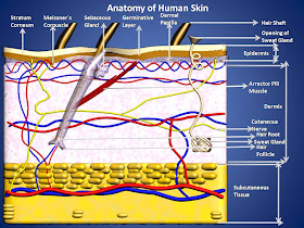
3D Picture of Human skin Created By Me (Manash Kundu)


SKIN:
The skin is the largest organ in the body and has a surface area of about 1.5 to 2 m square in adults and it contains glands, hair and nails.There are two main layers:
1) Epidermis
2) Dermis
Between the skin and underlying structures is a layer of subcutaneous fat.


3D Page of Human Skin in My Biology-World Software
 

Epidermis:
The epidermis is the most superficial layer of the skin and is composed of stratified keratinised squamous epithelium, which varies in thickness is different parts of the body.It is thickest on palms of the hands and soles of the feet.There are no blood vessels or nerve endings in the epidermis, but its deeper layers are bathed in interstitial fluid from the dermis, which provides oxygen and nutrients and drained away as lymph.
      
There are several layers(strata) of cells in the epidermis which extend from the deepest germinative layer to the surface stratum corneum ( a thick horny layer). The cells on the surface are flat, thin, non nucleated,dead cells or squames, in which the cytoplasm has been replaced by the fibrous protein keratin.These cells are constantly being rubbed off and replaced by cells that originated in the germinative layer and have undergone gradual change as they progressed towards
the surface.Complete replacement of the epidermis takes about a month.The maintenance of healty epidermis depends upon 3 process being synchronised:

1) Desquamation(shedding) of the keratinised cells from the surface.
2) Effective keratinisation of cells approaching the surface.
3) Continual cell division in the deeper layers with newly formed cells being pushed to the surface.

Hairs, secretions from sebaceous glands and ducts of sweat glands pass through the epidermis to reach the surface.
       
The surface of the epidermis is ridged by projections of cells in the dermis called papillae. The pattern of ridges is different in every individual and the impression made by them is the fingerprint.The downward projections of the germinative layer between the papillae are believed to aid nutrition of epidermal cells and stabilise the 2 layers, preventing damage due to shearing forces.Blister develop when trauma causes separation of the dermis and epidermis and epidermis and serous fluid collects between the 2 layers.Skin colour is affected by various factors:-

Melanin:
A dark pigment derived from the amino acid tyrosine and secreted by melanocytes in the deep germinative layer, is absorbed by surrounding epithelial cells.The amount of genetically determined and varies between different parts of the body, between people of same ethnic origin and between ethnic groups.The number of melanocytes is fairly constant so the differences in colour depend on the amount of melanin secreted.It protects the skin fromthe harmful effects of sunlight.Exposure to sunlight promotes synthesis of melanin.

The percentage saturation of haemoglobin and the amount of blood circulating in the dermis give white skin its pink colour.

Excessive level of bile pigments in blood and carotenes in subcutaneous fat give the skin a yellowish colour.

Dermis:
The dermis is tough and elastic.It is formed from connective tissue and the matrix contains collagen fibres interlaced with elastic fibres. Rupture of elastic fibres occur when the skin is overstreched, resulting in permanent striae, or strech marks that may be found in pregnancy and obesity.Collagen fibres bind water and give the skin its tensile strength,but as its ability declines with age,wrinkle develop.Fibroblasts, macrophages and mast cells found in the dermis.Underlying its deepest layer there is a  areolar tissue and varying amounts of adipose tissue.The structure in the dermis are:
Lymph vessels:
Sensory nerve endings:
Sweat glands and their ducts:
Hairs,arrector pili muscles and sebaceous glands:


3D Picture of Human skin Created By Me (Manash Kundu)
 
Sensory nerve endings:

Sensory receptor(specialised nerve endings) sensative to touch, temperature, pressure and pain are widely distributed in the dermis. Incoming stimuli activate different types of sensory receptors. The skin is  an important sensory organ through which individual receive information about their environment. Nerve impulses generated in the sensory receptors in the dermis, are conveyed to the spinal cord by sensory nerves, then to the sensory area of the cerebrum
where the sensations are perceived.
Sensory Receptors:                                                                             Stimulus:
Meessner`s corpuscle                                                                         Light pressure
Pacinian Corpuscle                                                                             Deep pressure
Free nerve ending                                                                                Pain

Sweat Glands:
Sweat glands are widely distributed throughtout the skin and  are most numerous in the palms of the hands, soles of the feet axillae and groins.They are composed of epithelial cells.The bodies of the glands lie coiled in the subcutaneous tissue. Some ducts open onto the skin surface at tiny depressions, or pores and others open into hair follicles do not become active until puberty.In the axilla they secrete an colourless milky fluid which, if decomposed by surface microbes, causes an unpleasant odour.The functions of this secretion are not known. Sweat glands are stimulated by sympathetic nerves in response to raised body temperature and fear.

Hairs:
These are formed by a down-growth of epidermal cells into the dermis or subcutaneous tissue, called hair follicles. At the base of the follicle is a cluster of cells called the bulb.The hair is formed by multiplication of cells of the bulband as they are pushed upwards ,away from their source of nutrition, the cell die and become keratinised.The part of the hair above the skin is the shaft and the remainder, the root.

Functions of the skin:


1) Protection:
Defence action barrier against:
Invasion by microbes:
Chemical:
Physical Agents e.g. Mild trauma,Ultraviolet light:
Dehydration:

2) Regulation of body temperature:

3) Heat production:
 The Muscles:- Contraction of skeletal muscles produces a large amount heat and the more strenuous produces a large amount heat and the more strenuous the muscular exercise, the greater the heat produced.Shivering also involves skeletal muscle produced.

The liver is very metabolically active:-

The digestive organs:-  Produces heat during peristalsis and during the chemical reactions involved in digestion.

4) Heat Loss:

5) Formation of vitamin D:

6) Cutaneous Sensation:

7) Absorption:

8) Excretion:


3D Picture of Human Tongue Created By Me (Manash Kundu)
 

SENSE OF TASTE:
4 fundamental sesations of taste have been described -sweet, sour, bitter, and salt.This is probably an oversimplification
because perception varies widely and many tastes cannot be easily classified.However, some tastes consistently stimulate
taste buds in specific parts of the tongue:

1) Sweet and salty mainly at the tip.
2) Sour, at the sides.
3) Bitter, at the back.

The sense of taste triggers salivation and the secretion of gastric juice.It also has a protective function, e.g. when foul-tasting food is eaten, reflex gagging or vomiting may be induced.The sense of taste is impared when the mouth is dry.because substances can only be `tasted` when in solution.


3D Picture of Human Female Breast Created By Me (Manash Kundu)
 

BREASTS
The breasts or mammary glands are accessory glands of the female reproductive system. They exist also in the male, but in only a rudimentory form. In the female, the breasts are small and immature until puberty.Thereafter they grow and develop under the influence of oestrogen and progesterone.During pregnancy these hormones stimulates further growth.After the baby is born the hormone prolactine from the anterior pituitory stimulates the production of milk, and oxytocin from the posterior pituitory stimulates the release of milk in response to the stimulation of the nipple by the sucking baby, by a positive feedback mechanism.

Structure:
The mammary glands consist of glandular tissue, fibrous tissue and fatty tissue.Each breast consists of about 20 lobes of glandular tissue, each lobe being made up of a number of lobules that radiate around the nipple. The lobule consist of a cluster of alveoli that open into small ducts, and these unite to form large excretory ducts called lactiferous ducts. They form dilatations or reservoirs for milk. leading from each dilatation, or lactiferous sinus is anarrow duct that opens on to the surface of nipple.
    
The nipple is small conical eminence at the centre of the surrounded by the a pigmented area, the areola.On the surface of areola are numerous sebaceous glands , which lubricate the nipple during lactation.

Function:
The mammary glands are only active during late pregnancy and after childbirth, when they produce milk lactation.Lactation is stimulated by the hormone prolactin.


3D Picture of Human Tear Gland Created By Me (Manash Kundu)
 

LACRIMAL APPARATUS (Tear Gland) :
For each eye this consists of:

1 Lacrimal gland  and its ducts.
2 lacrimal canaliculi.
1 lacrimal sac
1 Nasolacrimal duct.

The lacrimal glands are exocrine glands situated in recesses in the frontal bones on the lateral aspect  of each eye just behind the supraorbital margin. Each gland is approximately the size and shape of an almond, and is composed of secretory epithelial cells.The glands secrete tears composed of water, mineral salts, antibodies, and lysozyme,a bactericidal enzyme.
 
The tears leave the lacrimal gland by several small ducts and pass over the front of the eye under the lids towards the medial canthus where they drain into the 2 lacrimal canaliculi; the opening of each is called the punctum.The 2 canaliculi lie one above the other, separated by small red body, the caruncle.The tears then drain into the lacrimal sac,which is the upper expanded end of nasolacrimal duct.This is the membranous canal approsimately 2 cm long, extending from the lower part of the lacrimal sac to the nasal cavity, opening at the level of inferior concha. Normally the rate of secretion of tears keep pace with the rate of drainage.When a foreign body or other irritant enters the eye the secretion of tears is greatly increased and the conjunctival blood vessels dilate.Secretion of tears is greatly increased and the conjunctival blood vessels dilate. Secretion of tears is also increased in emotional states, e.g. crying, laughing.

Functions:
Washing away irritating materials, e.g. dust, grit.
The bacteriocidal enzyme lysozyme prevents microbial infection.
Its oiliness delays evaporation and prevents drying of the conjunctiva. 


3D Picture of Human Nose created By Me (Manash Kundu)
 

OLFACTORY NERVES:

These are the sensory nerves of smell. they originate as specialised olfactory nerve endings(Chemoreceptor) in the mucous membrane of the roof of the nasal cavity above the superior nasal conchae. On each side of the nasal septum nerve fibres pass through the cribriform plate of the ethmoid bone to the olfactory bulb where interconnections and synapses occur. From the bulb, bundles of nerve fibres form the olfactory tract , which passes backwards to the olfactory area in the temporal lobe of the cerebral cortex in each hemisphere where the impulses are interpreted and odour perceived.

Physiology:
The human sense of smell is less acute than in other animals. Many animals secrete odorous chemicals called pheromones that play important part in chemical communication in, for example, territorial behavior, mating and bonding of mothers and their newborn offspring.The role of pheromones in human communication is unknown. All odorous materials give off volatile molecules which are carried into the nose with the inhaled air and even very low concentrations, when dissolved in mucus, stimulate the olfactory chemoreceptors.
    
The air entering the nose is warmed, and convection currents carry eddies of inspired air to the roof of the nasal cavity. Sniffing concentrates volatile molecules in the roof of the nose.This increases the number of olfactory receptors stimulated and thus the perception of smell.The sense of smell affect the appetite may improve and vice versa.When accopanied bt sight of food, and an appetising smell increases salivation and stimulates the digestive systems.The sense of smell may create long lasting memories, especially for distinctive odours, e.g. hospital smells,favourite or least-liked foods.


3D Picture of Human main Sense Organs Created By Me (Manash Kundu)


Human Ear : Auditory Organ of Hearing of Human Body


Human Ear Page of my Bio-World Software


EAR:
The ear is the organ of hearing.It is supplied by 8th cranial nerve, i.e. the cochlear part of the vestibulo cochlear nerve, which is stimulated by vibrations caused by sound waves.
 

With the exception of the auricle(pinna), the structures that form the ear are encased within the petrous portion of the temporal bone.

Structure:
The ear is divided into 3 dictinct parts
1) Outer ear
2) Middle ear (Tympanic cavity)
3) Inner ear.


3D Picture of Different Parts of Human Ear created by me (Manash Kundu)


Outer ear:
The outer ear consists of the auricle(pinna) and the external acoustic meatus(auditory canal).

The auricle(pinna):

 
The auricle is expanded portion that projects from the side of the head.It is composed of fibroelastic cartilage covered with skin.It is deeply grooved and ridged; the most prominent outer ridge is the helix.
   
The lobule(earlobe) is the soft pliable part at the lower extremity, composed of fibrous and adipose tissue richly supplied with blood.

External acoustic meatus(Auditory Canal):

 
This is a slightly S-shaped tube about 2.5 cm long extending from the auricle to the tympanic membrane(eardrum).The lateral third is cartilaginous and the remainder is a canal in the temporal bone.The meatus is lined with skin continuous with that with the auricle.There are numerous ceruminous glands and hair follicles, with associated sebaceous glands. In the skin of lateral third.Ceruminous glands are modified sweat glands that secrete cerumen(Earwax), a sticky material containing lysozyme and immunoglobins.Foreign materials e.g. dust, insects and microbes, are prevented from reaching the tympanic membrane bu wax, hair and the curvature of meatus.Movement of temporomandibular joint during chewing and speaking `massage` the cartilaginous meatus , moving the wax towards the exterior.


3D Picture of Three Parts of The Human Ear created by me (Manash Kundu)
 

Middle ear(Tympanic Cavity):

This is an irregular-shaped air-filled cavity within the petous portion of the temporal bone.The cavity, its contents and the air sacs which open out of it are lined with either simple squamous or cuboidal epithelium.The lateral wall of the middle ear is formed by the tympanic membrane.The roof and floor are formed by the temporal bone.The posterior wall is formed by the temporal bone with openings leading to the mastoid antrum through which air passes to the air cells within the mastoid process.The median wall is a thin layer of temporal bone in which there are 2 openings:

  •      Oval window
  •      Round window 
The oval window is occluded by part of a small bone called the stapes and the round window, by a fine sheet of fibrous tissue.Air reaches the cavity through the pharyngotympanic (Auditory or Eustachian) tube, which extends from the nasopharynx.It is about 4 cm long and is lined with ciliated columner epithelium.The presence of air at atmospheric pressure on both sides of the tympanic membrane is maintained by the pharyngotympanic tube and enables the membrane to vibrate when sound waves strike it.The pharyngotympanic tube is normally closed but when there is unequal pressure across the tympanic membrane, e.g. at high altitude,it is opened by swallowing or yawning and the ears `pop`,equalising the pressure again.

Auditory Ossicles:

 
These are 3 very small bones that extend across the middle ear from the tympanic membrane to the oval window.They form a series of movable joints with each other and with the medial wall of the cavity at the oval window.They are named
according to their shapes.

The Malleus:

 
This is the lateral hammer-shaped bone. The handle is in contact with the tympanic membrane and the head forms a movable joint with the Incus.

The incus:
 

This is the middle anvil-shaped bone. Its body articulates with the malleus, the long process with stapes, and it is stabilised by the short process, fixed by fibrous tissue to the posterior wall of the tympanic cavity.

The stapes:
 

This is medial stirrup-shaped bone.Its head articulates with the incus and its footplate fits into the oval window. The 3 ossicles are held in position by fine ligaments.

3D Picture Different Parts of The Inner Ear created by me (Manash Kundu)


Inner ear:
The inner ear or labyrinth contains the organs of hearing and balance.It is described in 2 parts, the bony labyrinth and the membranous labyrinth.


Inner Ear
 

Bony Labyrinth:

 
This is a cavity within the temporal bone lined with periosteum. It is larger than,and encloses, the membranous labyrinth of the same shape that fits into it, like a tube within a tube.Between the bony and membranous labyrinth there is a layer of watery fluid called perilymph and within the membranous labyrinth there is a similarly watery fluid, endolymph.

The bony labyrinth consists of :

  • The vestibule.
  • The cochlea.
  • Three semicircular canals.
 
Vestibule:

 
This is the expanded part nearest the middle ear. It contains the oval and round windows in its lateral wall.

Membranous labyrinth:

 
This contains endolymph and lies within its bony counterpart. It comprises


  • The vestibule Which contain utricle and saccule
  • The cochlea.
  • Three semicircular canals.
Cochlea;
 
A cross section of the cochlea contains three compartments:


  • The scala vestibuli.
  • The scala media, or cochlear duct
  • The scala tympani. 
In the cross section the bony cochlea has 2 compartments containing perilymph;The scala vestibuli, which originates at the oval window,The compartments are continuous with each other the cochlear duct is the part of the membranous labyrinth and its triangular in shape. On the basilar membrane or the base of the triangle there are supporting cells and specialised hair cells containing auditory receptors. These cells form the spiral organs(of corti).Then sensory organ that responds to vibration by initiating nerve impulses that are then perceived as hearing by the brain. The auditory receptors are  dendrites of the efferent nerves that combine forming  the cochlear part of the vestibocochlear nerve (8Th cranial nerve), which passes through a foramen in the temporal lobe of the cerebrum.

VESTIBULOCOCHLEAR NERVES (AUDITORY)(SENSORY):

 
These nerves are composed of two distinct sets of fibres,vestibular nerves and cochlear nerves.
The Vestibular nerves arise from the semicircular canals of the inner ear and convey impulses to the cerebellum. they are associated  with the maintenance of posture and balance.
The Cochlear nerves originate in the spiral organ in the inner ear and convey impulses to the hearing area in the cerebral cortex where sound is perceived.


Detail Picture of Different Parts of The Ear
 

Semicircular canals:

 
These are 3 tubes arranged so that one is situated in each of the 3 planes of space.They are continuous with the vestibule


3D Picture of Three Parts of The Human Ear created by me (Manash Kundu)


PHYSIOLOGY OF HEARING:
Every sound produces sound waves or vibration in the air, which travel at about 332 metres per sec.The auricle, because of its shape, collects and concentrates the waves and directs  them along the external acoustic meatus causing the Tympanic membrane to vibrate.Tympanic membrane vibrations are transmitted and amplified through the middle ear by movement of the ossicles.At their medial end the footplate of the shape rocks to and fro in the oval window, setting up fluid waves in the perilymph of the scala vestibuli.Some of the force of these waves is transmitted along the length of the scala-vestibuli and scala tympany, but most of the pressure is transmitted into the cochlear duct.This causes a corresponding wave motion in the endolymph,resulting in vibration of the basilar membrane and stimulation of the auditory receptors in the hair cells of the spiral organ.


Physiology or Process of Hearing


The nerve impulse generated pass to the brain in the cochlear of the vestibo cochlear nerve(8th cranial nerve).The fluid wave is finally expended into the middle ear by vibration of the membrane of the round window. The 8th cranial nerve transmit the impulses to the auditory nuclei in the medulla ,where they synapse before they are conducted to the auditory area in the temporal lobe of the cerebrum.Because some fibres cross over in the medulla and others remain on the same side, the left and right auditory areas of the cerebrum receives impulses from both ears.
         
Sound waves have the properties of pitch and volume, or intensity.Pitch is determined by the frequency of the sound waves and is measured in Hertz(Hz).Sounds of different frequencies stimulate the basilar membrane at different places along its length. allowing discrimination of pitch.


Human Ear Post

Human Eyes : Visual opening and Major Sense Organ Of Human Body

Eye Page In Bio-World Software

STRUCTURE OF EYE:

Eye within the Eye Sockets in The Human Skull

 
There are 3 layers of tissue in the walls of the eye. They are:
1) The outer fibrous layer: Sclera and cornea.
2) The middle vascular layer or Uveal tract: choroid,ciliary body and iris.
3) The inner nervous tissue layer: retina.


3D picture of Different Captioned Parts of Human Eye

 Retina:
 

Different Cells Within The Eye Retina


This is the innermost layer of the wall of the eye.it is extremely delicate structure and is well adapted for stimulation by light rays.It is composed of several layer of nerve cell bodies and their axons, lying on the pigmented layer of epithelial cells which attach it to the choroid.The light sensitive layer consists of sensory receptor cell: Rods and cones.

The retina lines about three-quarters of the eyeball and thickest at the back., near the center of the posterior part is the macula lutea or yellow spot.In the center of the yellow spot is a little depression called the fovea centralis,Consisting of only cones.
        
The rods and cones contain photosensative pigments that convert light rays into nerve impulses. The small area of retina where the optic nerve leaves the eye is the optic disc of blind spot.It has no light sensitive cells.  


Different Parts of Human Eye
 

Sclera and cornea:

The sclera. or white of the eye, forms the outermost layer of the posterior and lateral aspects of the eyeball and is continuous anteriorly with the transparent cornea.It consists of a firm fibrous membrane that maintains the shape of the eye and gives attachment to the extrinsic muscles of the eye.
     
Anteriorly the sclera continues as a clear tranparent epeithelial membrane, the cornea, light rays pass through the  cornea to reach the retina. The cornea is convex anteriorly and is involved in refracting(Bending) light rays to focus them on the retina.


3D picture of Different Captioned Parts of Human Eye
 

lens:
The lens is highly elastic circular biconvex body, lying immidiately behind the pupil.It consist of fibres enclosed within a capsule and it is suspended from the ciliary body by the suspensory ligaments.When the ciliary muscle contracts, it moves forward,releasing its pull on the lens, increasing its thickness.The nearer is the object being viewed, the thicker the lens becomes to allow focusing.
  
The lens refracts light rays reflected by objects in front of the eye.It is the only structure in the eye that can vary its refractory power,which is achieved by changing its thickness.

Iris:
This is the visible coloured of the eye and extend anteriorly from the ciliary body,lying behind the cornea and in front of the lens.It divides the anterior segment of the eye into anterior and posterior chambers which contain aqueous fluid secreted by ciliary body.It is a circular body composed of pigment cells and 2 layers of smooth muscle fibres, one circular and other radiating.In the center there is a aperture called the pupil.
     
The iris is supplied by parasympathetic and sympathetic nerves.Parasympathetic constrics the pupil and sympathetic stimulation dilates.
     
The colour of iris is genetically determined and depends on the number of pigment cells present.Albinos have no pigment cells and people with blue eyes have fewer than those with brown eyes.

Ciliary Body:
This is the anterior continuation of the choroid consisting of ciliary muscle(smooth muscle fibres) and secretory epithelial cells.It gives attachment to the suspensory ligament, which at its other end, is attached to the capsule enclosing the lens.Contraction and relaxation of the ciliary muscle changes the thickness of the lens, which bends light rays entering the eye to focus them on the retina.The epithelial cells secrete aqueous fluid into the anterior segment of the eye, e.g. the space between the lens and the cornea. The ciliary body is supplied by parasympathetic branches of the occulomotor nerve.Stimulation causes contraction of the ciliary muscle and accomodation of the eye.

Optic Nerves:
The fibres of the optic nerve originate in the retina and they converge to form the optic nerve about 0.5 cm to the nasal side of the macula lutea.The nerve pierces the choroid and sclera to pass backwards and medially through the orbital  cavity. It then passes through the optic foramen of the sphenoid bone, backwards and medially to meet the nerve from the other eye at the optic chiasma.

OPTIC NERVES (SENSORY):
These are the nerves of sense of sight.The fibres originate in the retinae of the eyes and they combine to form the optic nerves.They are directed backward and medially through the posterior part of the orbital cavity.They then pass through the optic foramina of the sphenoid bone into the cranial cavity and join at the optic chiasma.The nerves proceed back- wards as the optic tracts to the lateral geniculate bodies of the thalamus.Impulses pass from these to the centre for sight in the occipital lobes of the cerebrum and to the cerebellum.In the occipital lobe sight is perceived,and in the cerebellum the impulses from the eyes contribute to the maintaenance of balance,posture and orientation of the head in space.


Optic Nerve Tracts
 

Optic Tracts:
These are the pathways of the optic nerves, posterior to the optic chiasma.Each tract consists of the nasal fibres from the retina of one eye and the temporal fibres from the retina of the other.The optic tracts pass backwards to synapse with the nerve cells of the lateral geniculate body of the thalamus.From there the nerve fibres proceed backwards and medially as the optic radiation to terminate in the visual area of the cerebral cortex in the occipital lobes of the cerebrum.Other neurones originating in the lateral geniculate bodies convey impulses from the eyes to the cerebellum where, together with impulses from the semicircular canals of the ears and from the skeletal muscles and joints, they contribute to the maintenance of posture and balance.

Optic Chiasma:
This is situated immediately in front of above the pituitory gland, which is in the hypophyseal fossa of the sphenoid bone.In the optic chiasma the nerve fibres of the optic nerve from the nasal side of each retina cross over to the opposite side.The fibres from the temporal side do not cross but continue backwards on the same side. This crossing over provides both cerebral hemispheres with sensory input from the each eye.


Binocular Vision of Human Eye
 

BINOCULAR VISION:
Binocular or stereoscopic vision enables 3D view although each eye sees a scene slightly differently.The visual fields overlap in the middle but the left eye sees more on the left than can be seen by the other eye and vice versa. The images from the 2 eyes are fused in the cerebrum so that only one image is perceived.
 
Binocular vision provides a much more accurate assessment of one object relative to another, e.g. its distance, depths height and width.People with monocular vision may find it difficult, for example, to judge the speed and distance of an approaching vehicle.

PHYSIOLOGY OF SIGHT:
 

Different Types of Wave with Visible Spectrum of Light

Light wave travel at a speed of 300 000 km(1,86,000 miles) per sec. The light is reflected into the eyes by object within the the field of vision.White light is the combination of all the colours of the visual spectrum(Rainbow) i.e. red orange,yellow , green , blue ,indigo and violet.This is demonstrated by passing white light through the glass prism which bends the rays of the different colours to a greater or lesser extent, depending on their wavelengths.Red light has the longest wavelengths .Red light has the longest wavelength and violet the shortest.
   
The range of colour is the spectrum of visible light. In a rainbow, while light from the sum is broken up by raindrops, which act as prisms and reflectors.


Eye Muscles of Human
 

MUSCLES OF EYES:
These include the muscles of the eyelids and those that move the eyeballs. The eyeball is moved 6 extrinsic muscles, attached at one end to the eyeball and at the other to the walls of the orbital cavity. There are 4 straight muscles and 2 oblique muscles.

Moving the eyes to took in a particular direction is under voluntry control, but coordination of movement, needed for convergence and accomodation to near or distant vision, is under autonomic(involuntry) control.Movements of the eyes resulting from the action of these muscles.


                                                    3D Animation Showing Path of Image Ray into the Eye.


Wednesday, 19 September 2012

Vishwakarma - Lord of Architecture: Ancient Hindu God of Engineering


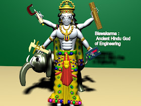
3D Picture of Lord Vishwakarma Created by Me (Manash Kundu)

Vishwakarma - Lord of Architecture: Ancient Hindu God of Engineering:

Vishwakarma is the presiding deity of all craftsmen and architects. Son of Brahma, he is the divine draftsman of the whole universe, and the official builder of all the gods' palaces. Vishwakarma is also the designer of all the flying chariots of the gods, and all their weapons.


Picture of multi-handed and multi-headed Ultimate form of Vishwakarma


people related to engineers types of work they celebrate bithday of Vishwakarma  and large number of people go to temple for worship Lord Vishwakarma. Because load Vishwakarma is know as a God of style and creation. People who have work related to creation or engineer  they don’t do any type work on the day, every carpenters, goldsmiths, blacksmiths, masons and all those people who have skilled work they pay proper worship to lord Vishwakarma. it s believed by the people that all types of art come from lord Vishwakarma. In the Rig Veda, Lord Vishwa Karma is the lovely engineer of the total world. He is the embodiment of the original power that welds world and heaven jointly. Hindu citizens say as Lord Vishwa luck is the son of Brahma and is the architect of all gods’ palaces. He is also known as God of manufacture.


3D Imaginary picture of Pushpak Vimana


Lord Vishwakarma uses Pushpak Vimaan a rapid chariot. His creations number of worldly spaces and weapons. He is the architect and builder of Dwarka, the place where Load Krishna live. He also creates town of Indraprastha and Hastinapur. Sudarsana Chakra and Agneyastra as well are also amongst his huge creations. In fact, legendary Golden Lanka (Sri Lanka) was also built by Lord VISHWAKARMA.


3D Picture of Lord Vishwakarma Created by Me (Manash Kundu)

Lord Vishwakarma decorated white and has a club in his wrists as well as holds a book, a water-pot, a noose and craftsman’s tools in his left hand. He is the revealer of the science of design and workings. Lord Vishwa Karma is called as Sthapatya Veda.

The Mahabharata describes him as "The lord of the arts, executor of a thousand handicrafts, the carpenter of the gods, the most eminent of artisans, the fashioner of all ornaments ... and a great and immortal god." He has four hands, wears a crown, loads of gold jewelry, and holds a water-pot, a book, a noose and craftsman's tools in his hands.


Lord VISHWAKARMA is described as the God of carpenters, goldsmiths, blacksmiths, masons and all those people who are skilled


Lord VISHWAKARMA with multi-hand is accepted by every Indian as the creator of Universe. Lord VISHWAKARMA is described as the God of carpenters, goldsmiths, blacksmiths, masons and all those people who are skilled. Lord VISHWAKARMA is as great as Lord BRAHMA, VISHNU, MAHESH because all scriptures concerned with Hindu religion contains episodes of his glorious contributions.

In the Vedas:

Vishwakarma is visualized as the Ultimate reality in the Rig Veda, from whose navel all visible things emanate. The same imagery is seen in Yajurveda purusha sukta, where the divine smith Tvastar originates from Vishwakarma.This concept developed later in the puranic period as Padmanabha. As he is invoked as AJA the unborn , he is the primordial personification of the upanishadic abstract concept Parabrahman.


Pancha Mukha Virt Vishwakarma


In the Vedic period the term first appeared as an epithet of Indra, Surya, and Agni. In that time Brahma might have been hidden in the concept of Vastospati or Brahmanaspati. In the last phase of vedic period and during the growth of monotheism, the top position of God was gradually assigned to Vishwakarma who was perceived as a hotar, a universal father, the creator and an all seeing god who has faces, eyes and feet on every side; and who produced all the Heavenly, Earthly and other Celestial realms and preserves them through the exercise of his arms and wings

Vishwakarma Puja:


Hindus widely regard Vishwakarma as the god of architecture and engineering, and September 16 or 17 every year is celebrated as Vishwakarma Puja — a resolution time for workers and craftsmen to increase productivity and gain divine inspiration for creating novel products. This ritual usually takes place within the factory premises or shop floor, and the otherwise mundane workshops come alive with fiesta. Vishwakarma Puja is also associated with the buoyant custom of flying kites. This occasion in a way also marks the start of the festive season that culminates in Diwali. 


Vishwakarma Puja is also associated with the buoyant custom of flying kites


Vishwakarma is known as the divine engineer of the world. As a mark of reverence he is not only worshipped by the engineering and architectural community but also by all professionals. It is customary for craftsmen to worship their tools in his name.

Like other gods, Vishwakarma is attributed a putative birthday by the Hindu religion. 


Vishwakarma Statue  at Mandi ,India

Vishwakarma Temple,Mandi

The more philosophical minded argue that it is impossible for the original Creator of everything to be born on a particular day. It is a contradiction in terms since that presupposes another creator for Vishwakarma. The Vishwakarma Puja is celebrated in all parts of India, especially in Assam, West Bengal, Rajasthan ,Uttarakhand (Dehardun).

Vishwakarma Idol in West Bengal


Vishwakarma's Architectural Wonders:

 
Hindu mythology is full of Vishwakarma's many architectural wonders. Through the four 'yugas', he had built several towns and palaces for the gods. In "Satya yuga", he built the Swarg Loke, or heaven, the abode of the gods and demigods where Lord Indra rules. Vishwakarma then built the 'Sone ki Lanka' in "Treta yuga", the city of Dwarka in "Dwapar yuga", and Hastinapur  in the "Kali yuga".

Sone Ki Lanka' or Golden Lanka:


Imaginary 3D Picture of Golden Lanka of Ravana


According to Hindu mythology, 'Sone ki Lanka' or Golden Lanka was the place where the demon king Ravana dwelled in the "Treta yuga." As we read in the epic story Ramayana, this was also the place where Ravana kept Sita, Lord Ram's wife as a hostage.

There is also a story behind the construction of Golden Lanka. When Lord Shiva married Parvati, he asked Vishwakarma to build a beautiful palace for them to reside. Vishwakarma put up a palace made of gold! For the housewarming ceremony, Shiva invited the wise Ravana to perform the "Grihapravesh" ritual. After the sacred ceremony when Shiva asked Ravana to ask anything in return as "Dakshina", Ravana, overwhelmed with the beauty and grandeur of the palace, asked Shiva for the golden palace itself! Shiva was obliged to accede to Ravana's wish, and the Golden Lanka became Ravana's palace.

Dwarka:


Dwarka Picture


Among the many mythical towns Viswakarma built is Dwarka, the capital of Lord Krishna. During the time of the Mahabharata, Lord Krishna is said to have lived in Dwarka, and made it his "Karma Bhoomi" or center of operation. That is why this place in nothern India has become a well known pilgrimage for the Hindus.