 |
| Eye Page In Bio-World Software |
STRUCTURE OF EYE:
 |
| Eye within the Eye Sockets in The Human Skull |
There are 3 layers of tissue in the walls of the eye. They are:
1) The outer fibrous layer: Sclera and cornea.
2) The middle vascular layer or Uveal tract: choroid,ciliary body and iris.
3) The inner nervous tissue layer: retina.
 |
| 3D picture of Different Captioned Parts of Human Eye |
Retina:
 |
| Different Cells Within The Eye Retina |
This is the innermost layer of the wall of the eye.it is extremely delicate structure and is well adapted for stimulation by light rays.It is composed of several layer of nerve cell bodies and their axons, lying on the pigmented layer of epithelial cells which attach it to the choroid.The light sensitive layer consists of sensory receptor cell: Rods and cones.
The retina lines about three-quarters of the eyeball and thickest at the back., near the center of the posterior part is the macula lutea or yellow spot.In the center of the yellow spot is a little depression called the fovea centralis,Consisting of only cones.
The rods and cones contain photosensative pigments that convert light rays into nerve impulses. The small area of retina where the optic nerve leaves the eye is the optic disc of blind spot.It has no light sensitive cells.
 |
| Different Parts of Human Eye |
Sclera and cornea:
The sclera. or white of the eye, forms the outermost layer of the posterior and lateral aspects of the eyeball and is continuous anteriorly with the transparent cornea.It consists of a firm fibrous membrane that maintains the shape of the eye and gives attachment to the extrinsic muscles of the eye.
Anteriorly the sclera continues as a clear tranparent epeithelial membrane, the cornea, light rays pass through the cornea to reach the retina. The cornea is convex anteriorly and is involved in refracting(Bending) light rays to focus them on the retina.
|
| 3D picture of Different Captioned Parts of Human Eye |
lens:
The lens is highly elastic circular biconvex body, lying immidiately behind the pupil.It consist of fibres enclosed within a capsule and it is suspended from the ciliary body by the suspensory ligaments.When the ciliary muscle contracts, it moves forward,releasing its pull on the lens, increasing its thickness.The nearer is the object being viewed, the thicker the lens becomes to allow focusing.
The lens refracts light rays reflected by objects in front of the eye.It is the only structure in the eye that can vary its refractory power,which is achieved by changing its thickness.
Iris:
This is the visible coloured of the eye and extend anteriorly from the ciliary body,lying behind the cornea and in front of the lens.It divides the anterior segment of the eye into anterior and posterior chambers which contain aqueous fluid secreted by ciliary body.It is a circular body composed of pigment cells and 2 layers of smooth muscle fibres, one circular and other radiating.In the center there is a aperture called the pupil.
The iris is supplied by parasympathetic and sympathetic nerves.Parasympathetic constrics the pupil and sympathetic stimulation dilates.
The colour of iris is genetically determined and depends on the number of pigment cells present.Albinos have no pigment cells and people with blue eyes have fewer than those with brown eyes.
Ciliary Body:
This is the anterior continuation of the choroid consisting of ciliary muscle(smooth muscle fibres) and secretory epithelial cells.It gives attachment to the suspensory ligament, which at its other end, is attached to the capsule enclosing the lens.Contraction and relaxation of the ciliary muscle changes the thickness of the lens, which bends light rays entering the eye to focus them on the retina.The epithelial cells secrete aqueous fluid into the anterior segment of the eye, e.g. the space between the lens and the cornea. The ciliary body is supplied by parasympathetic branches of the occulomotor nerve.Stimulation causes contraction of the ciliary muscle and accomodation of the eye.
Optic Nerves:
The fibres of the optic nerve originate in the retina and they converge to form the optic nerve about 0.5 cm to the nasal side of the macula lutea.The nerve pierces the choroid and sclera to pass backwards and medially through the orbital cavity. It then passes through the optic foramen of the sphenoid bone, backwards and medially to meet the nerve from the other eye at the optic chiasma.
OPTIC NERVES (SENSORY):
These are the nerves of sense of sight.The fibres originate in the retinae of the eyes and they combine to form the optic nerves.They are directed backward and medially through the posterior part of the orbital cavity.They then pass through the optic foramina of the sphenoid bone into the cranial cavity and join at the optic chiasma.The nerves proceed back- wards as the optic tracts to the lateral geniculate bodies of the thalamus.Impulses pass from these to the centre for sight in the occipital lobes of the cerebrum and to the cerebellum.In the occipital lobe sight is perceived,and in the cerebellum the impulses from the eyes contribute to the maintaenance of balance,posture and orientation of the head in space.
 |
| Optic Nerve Tracts |
Optic Tracts:
These are the pathways of the optic nerves, posterior to the optic chiasma.Each tract consists of the nasal fibres from the retina of one eye and the temporal fibres from the retina of the other.The optic tracts pass backwards to synapse with the nerve cells of the lateral geniculate body of the thalamus.From there the nerve fibres proceed backwards and medially as the optic radiation to terminate in the visual area of the cerebral cortex in the occipital lobes of the cerebrum.Other neurones originating in the lateral geniculate bodies convey impulses from the eyes to the cerebellum where, together with impulses from the semicircular canals of the ears and from the skeletal muscles and joints, they contribute to the maintenance of posture and balance.
Optic Chiasma:
This is situated immediately in front of above the pituitory gland, which is in the hypophyseal fossa of the sphenoid bone.In the optic chiasma the nerve fibres of the optic nerve from the nasal side of each retina cross over to the opposite side.The fibres from the temporal side do not cross but continue backwards on the same side. This crossing over provides both cerebral hemispheres with sensory input from the each eye.
 |
| Binocular Vision of Human Eye |
BINOCULAR VISION:
Binocular or stereoscopic vision enables 3D view although each eye sees a scene slightly differently.The visual fields overlap in the middle but the left eye sees more on the left than can be seen by the other eye and vice versa. The images from the 2 eyes are fused in the cerebrum so that only one image is perceived.
Binocular vision provides a much more accurate assessment of one object relative to another, e.g. its distance, depths height and width.People with monocular vision may find it difficult, for example, to judge the speed and distance of an approaching vehicle.
PHYSIOLOGY OF SIGHT:
 |
| Different Types of Wave with Visible Spectrum of Light |
Light wave travel at a speed of 300 000 km(1,86,000 miles) per sec. The light is reflected into the eyes by object within the the field of vision.White light is the combination of all the colours of the visual spectrum(Rainbow) i.e. red orange,yellow , green , blue ,indigo and violet.This is demonstrated by passing white light through the glass prism which bends the rays of the different colours to a greater or lesser extent, depending on their wavelengths.Red light has the longest wavelengths .Red light has the longest wavelength and violet the shortest.
The range of colour is the spectrum of visible light. In a rainbow, while light from the sum is broken up by raindrops, which act as prisms and reflectors.
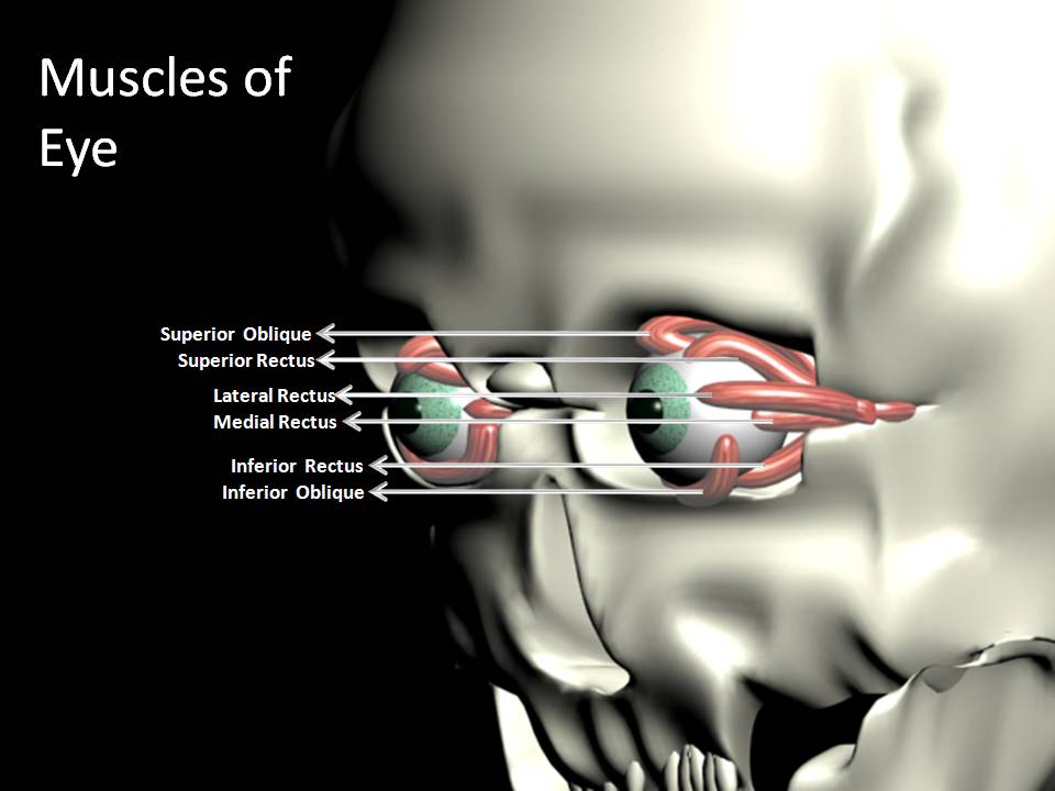 |
| Eye Muscles of Human |
MUSCLES OF EYES:
These include the muscles of the eyelids and those that move the eyeballs. The eyeball is moved 6 extrinsic muscles, attached at one end to the eyeball and at the other to the walls of the orbital cavity. There are 4 straight muscles and 2 oblique muscles.
Moving the eyes to took in a particular direction is under voluntry control, but coordination of movement, needed for convergence and accomodation to near or distant vision, is under autonomic(involuntry) control.Movements of the eyes resulting from the action of these muscles.
3D Animation Showing Path of Image Ray into the Eye.
No comments:
Post a Comment